Responsible: Camilla Baratto
Introduction
The most recent research activities concern the application of photoluminescence (PL) or Raman spectroscopy to materials of technological interest (oxides, semiconductors [1,2,3], 2D materials [4]), to fuel and bio-based plastics, to organic materials (plants [5], or cells [6]). Aspects covered include structural and dynamic characterization of crystalline materials, dimensional effects in nanocrystalline systems, crystallization processes and phase transformations, the recognition of tissue mineralisation and the study of chemometric techniques applied to spectroscopy.
Instrumentation and allowed configurations
Horiba modular micro-Raman allows to perform micro spectroscopy PL and Raman. With UV excitation (He-Cd 325 nm laser), PL measurements can be performed focusing with 50X LWD and 20X UV lenses in a controlled environment. Confocal Raman measurements are available with excitation at 442nm and 532nm. Horiba LabRam HR micro-Raman allows Raman spectroscopy with light excitation at 633 nm, 785 nm and 20X, 100X objectives.

Operando spectroscopy: thanks to the integration of a certified cylinder flow system and the use of a Linkam thermostatic cell, measurements can be made in a controlled environment between 77k and 873K.
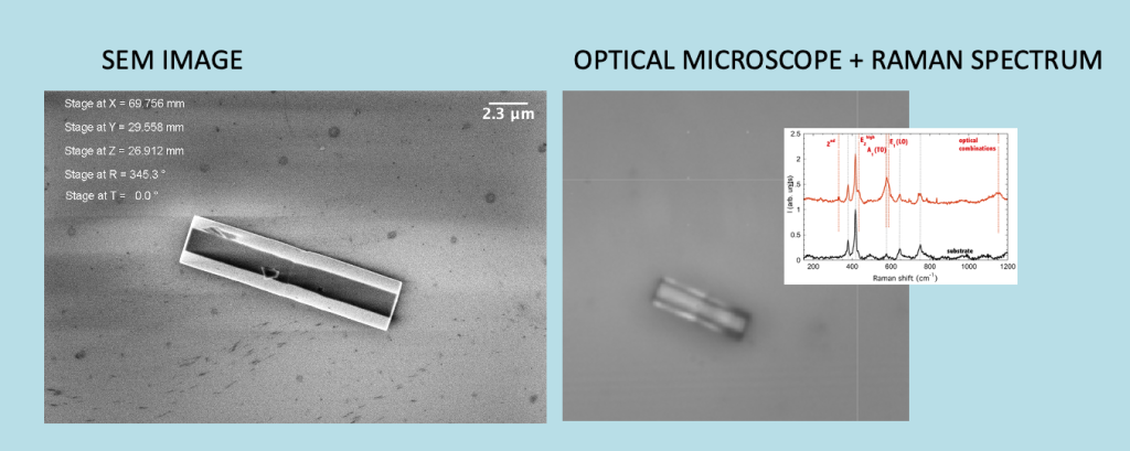
Correlative microscopy measurements between scanning microscopy and spectroscopy are also possible, allowing the morphological information of nano, micro or macro structures to be linked with information of a chemical-compositional nature of the sample.
Linked publications:
- L. Pandolfi, N. Miotti, G. Faglia, C. Pennacchio, A. Ponzoni, M. Ciuffo, S. Palmano, M. Schillaci, E. Gobbi, M. Turina, C. Baratto, Non-invasive Raman spectroscopy for monitoring metabolite changes in tomato plants infected by phytoplasma”, Anal. Methods 2025, 17, 5062.
- C. Baratto, M. Gandolfi, A. Tognazzi, P. Franceschini, G. Ambrosio, B. Li, R. C. Morales, D. De Ceglia, A. C. Cino, D. N. Neshev, “Electromagnetic wave sensors Optical Limiting Sensor Based on Multilayer Optimization of Ag/VO2 Phase Change Material,” vol. 7, no. 9, pp. 7–9, 2023, doi: 10.1109/LSENS.2023.3300801.
- F. Rigoni, D. Zappa, C. Baratto, G. Faglia, and E. Comini, “Single ZnO Nanowire for Electrical and Optical NO 2 Gas Sensing: Origin of Reversible and Irreversible Gas Effects Investigated by Photoluminescence Spectroscopy,” no. 2, 2024, doi: 10.1021/acssensors.4c00901.
- C. Baratto, G. Ambrosio, G. Faglia, and M. Turina, “Early detection of esca disease in asymptomatic vines by Raman Spectroscopy,” IEEE Sens. J., vol. 22, no. 23, p. 1, 2022, doi: 10.1109/JSEN.2022.3211616
- G. Ambrosio, G. Faglia, S. Tagliabue, and C. Baratto, “Study of the degradation of biobased plastic after stress tests in water,” Coatings, vol. 11, no. 11, pp. 1–18, 2021, doi: 10.3390/coatings11111330.
- C. Baratto, “Growth and properties of ZnO nanorods by RF-sputtering for detection of toxic gases,” Rsc Adv., vol. 8, no. 56, pp. 32038–32043, 2018, doi: 10.1039/c8ra05357j.
- C. Baratto, E. Comini, M. Ferroni, G. Faglia, and G. Sberveglieri, “Plasma-induced enhancement of UV photoluminescence in ZnO nanowires,” CrystEngComm, vol. 15, no. 39, pp. 7981–7986, 2013, doi: 10.1039/c3c
- C. Baratto et al., “Luminescence response of ZnO nanowires to gas adsorption,” Sensors Actuators, B Chem., vol. 140, no. 2, pp. 461–466, 2009, doi: 10.1016/j.snb.2009.05.018.
- G. Faglia, M. Ferroni, T. T. le Dang, M. Donarelli, F. Rigoni, and C. Baratto, “Vertically coupling ZnO nanorods onto MoS2 flakes for optical gas sensing,” Chemosensors, vol. 8, no. 1, pp. 1–12, 2020, doi: 10.3390/chemosensors8010019
- F. Re et al., “Mineralization of 3D osteogenic model based on gelatin-dextran hybrid hydrogel scaffold bioengineered with mesenchymal stromal cells: A multiparametric evaluation,” Materials (Basel)., vol. 14, no. 14, pp. 1–23, 2021, doi: 10.3390/ma14143852.